Welcome to the Amira-Avizo Software Use Case Gallery
Below you will find a collection of use cases of our 3D data visualization and analysis software. These use cases include scientific publications, articles, papers, posters, presentations or even videos that show how Amira-Avizo Software is used to address various scientific and industrial research topics.
Use the Domain selector to filter by main application area, and use the Search box to enter keywords related to specific topics you are interested in.
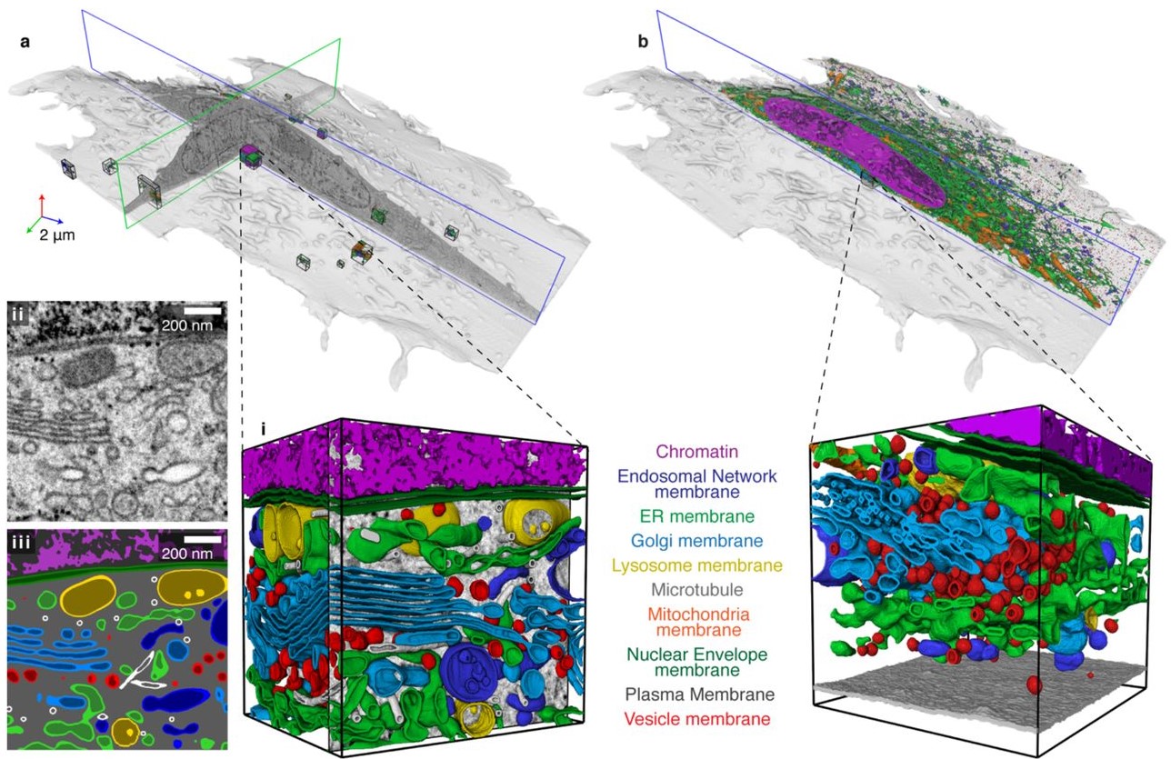
Automatic whole cell organelle segmentation in volumetric electron microscopy
Cells contain hundreds of different organelle and macromolecular assemblies intricately organized relative to each other to meet any cellular demands. Obtaining a complete understanding of their organization is challenging and requires nanometer-level, three-dimensional reconstruction of whole cells. Even then, the immense size of datasets and large number of structures to be characterized requires generalizable, automatic methods.
To meet this challenge, we developed an analy... Read more
Larissa Heinrich, Davis Bennett, David Ackerman, Woohyun Park, John Bogovic, View ORCID ProfileNils Eckstein, Alyson Petruncio, Jody Clements, C. Shan Xu, Jan Funke, Wyatt Korff, Harald F. Hess, Jennifer Lippincott-Schwartz, Stephan Saalfeld, Aubrey V. Weigel, COSEM Project Team
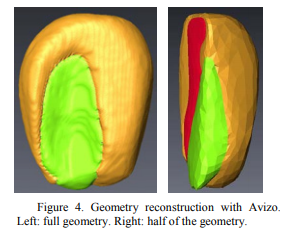
Drying of Corn Kernels: From Experimental Images to Multiscale Multiphysics Modeling
This work demonstrated the importance and feasibility of experimental image
to simulation workflow. The workflow is successfully applied to a food processing study, where multiphysics and multiscale modeling
based on 3D experimental image reconstruction contributes to the preservation of corn, one of the major food sources for the world population.
Corn kernels have a complex structure as they are composed of a pericarp layer outside and contain hard and soft endosperm and ... Read more
Pawan S. Takhar, and Shuang Zhang
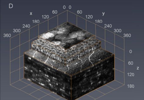
Cellular in vivo 3D imaging of the cornea by confocal laser scanning microscopy
We present an in vivo confocal laser scanning microscopy based method for large 3D reconstruction of the cornea on a cellular level with cropped volume sizes up to 266 x 286x 396 µm3.
The microscope objective used is equipped with a piezo actuator for automated, fast and precise closed-loop focal plane control. Furthermore, we present a novel concave surface contact cap, which significantly reduces eye movements by up to 87%, hence increasing the overlapping image area of the whole st... Read more
Sebastian Bohn, Karsten Sperlich, Stephan Allgeier, Andreas Bartschat, Ruby Prakasam, Klaus-martin Reichert, Heinrich Stolz, Rudolf Guthoff, Ralf Mikut, Bernd Köhler, and Oliver Stachs
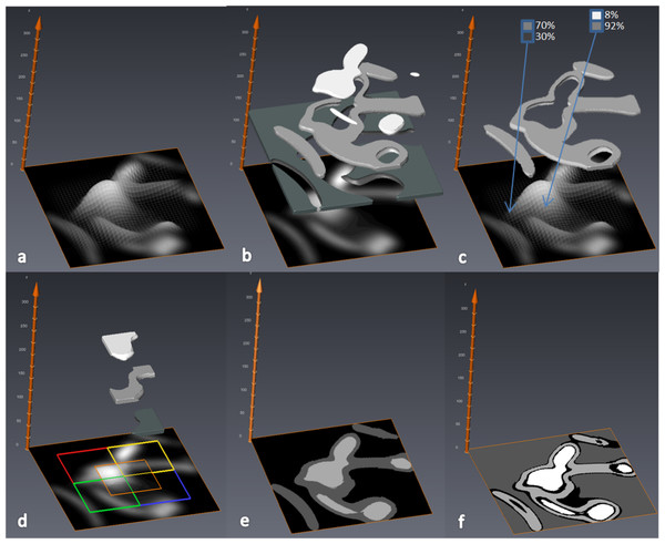
MIA-Clustering: a novel method for segmentation of paleontological material
Paleontological research increasingly uses high-resolution micro-computed tomography (μCT) to study the inner architecture of modern and fossil bone material to answer important questions regarding vertebrate evolution. This non-destructive method allows for the measurement of otherwise inaccessible morphology. Digital measurement is predicated on the accurate segmentation of modern or fossilized bone from other structures imaged in μCT scans, as errors in segmentation can result in inaccur... Read more
Christopher J. Dunmore, Gert Wollny, Matthew M. Skinner